Home

výskyt fond nerovnosť true lv aneurysm otto echo experiment neozbrojený kontakt

Cardiac Ultrasound (Echocardiography) Made Easy: Step-By-Step Guide - POCUS  101
Cardiac Ultrasound (Echocardiography) Made Easy: Step-By-Step Guide - POCUS 101

A rare case of true and pseudoaneurysm of left ventricular wall and  incremental value of myocardial contrast Kalra GS, Tandon R, Singh B, Mohan  B - J Indian Acad Echocardiogr Cardiovasc Imaging
A rare case of true and pseudoaneurysm of left ventricular wall and incremental value of myocardial contrast Kalra GS, Tandon R, Singh B, Mohan B - J Indian Acad Echocardiogr Cardiovasc Imaging
Echocardiography - ScienceDirect

Cureus | A Growing Two-Decade-Old True Left Ventricular Aneurysm: A Case  Report | Article
Cureus | A Growing Two-Decade-Old True Left Ventricular Aneurysm: A Case Report | Article

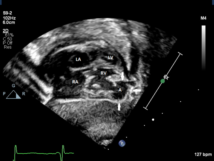
Congenital right ventricular aneurysm with characteristics of a  pseudoaneurysm | Cardiology in the Young | Cambridge Core
Congenital right ventricular aneurysm with characteristics of a pseudoaneurysm | Cardiology in the Young | Cambridge Core

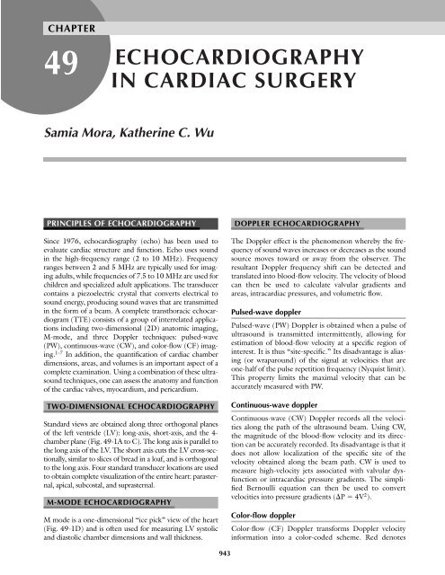
ECHOCARDIOGRAPHY IN CARDIAC SURGERY - TSDA

Transthoracic echocardiographic image of left ventricular septal... |  Download Scientific Diagram
Transthoracic echocardiographic image of left ventricular septal... | Download Scientific Diagram

Echo assessment of coronary artery disease

Figure, LV aneurysm. Image courtesy S Bhimji MD] - StatPearls - NCBI  Bookshelf
Figure, LV aneurysm. Image courtesy S Bhimji MD] - StatPearls - NCBI Bookshelf

Anatomical and physiological complications related to left ventricular  apical aneurysm - Stoodley - 2017 - Sonography - Wiley Online Library
Anatomical and physiological complications related to left ventricular apical aneurysm - Stoodley - 2017 - Sonography - Wiley Online Library

Medicowesome: LV aneurysm: Difference between true LV aneurysm and LV  pseudoaneurysm
Medicowesome: LV aneurysm: Difference between true LV aneurysm and LV pseudoaneurysm

Giant apical LV aneurysm - 3D TTE

e 2-Dimensional Echo in apical 4 chamber view showing large LV apical... |  Download Scientific Diagram
e 2-Dimensional Echo in apical 4 chamber view showing large LV apical... | Download Scientific Diagram

Transesophageal echocardiography. Two-chamber view. LV pseudoaneurysm.... |  Download Scientific Diagram
Transesophageal echocardiography. Two-chamber view. LV pseudoaneurysm.... | Download Scientific Diagram

Transthoracic Echocardiography: Beginner's Guide with Emphasis on Blind  Spots as Identified with CT and MRI | RadioGraphics
Transthoracic Echocardiography: Beginner's Guide with Emphasis on Blind Spots as Identified with CT and MRI | RadioGraphics

Traumatic left ventricular true aneurysm: echocardiographic, MRI, and  intraoperative images | Heart
Traumatic left ventricular true aneurysm: echocardiographic, MRI, and intraoperative images | Heart

Serial hemodynamic assessment using Doppler echocardiography in a fetus  with left ventricular aneurysm presented as fetal hydrops | Journal of  Perinatology
Serial hemodynamic assessment using Doppler echocardiography in a fetus with left ventricular aneurysm presented as fetal hydrops | Journal of Perinatology

Left ventricular apical aneurysm associated with normal coronary arteries  following cardiac surgery: Echocardiographic features and differential  diagnosis - ScienceDirect
Left ventricular apical aneurysm associated with normal coronary arteries following cardiac surgery: Echocardiographic features and differential diagnosis - ScienceDirect

Multimodality Imaging Demonstrating an Apical Variant Hypertrophic  Cardiomyopathy in an Uncommon Pentad - Ayman R. Fath, Clinton E. Jokerst,  Amro Aglan, Nawfal Mihyawi, Farouk Mookadam, 2020
Multimodality Imaging Demonstrating an Apical Variant Hypertrophic Cardiomyopathy in an Uncommon Pentad - Ayman R. Fath, Clinton E. Jokerst, Amro Aglan, Nawfal Mihyawi, Farouk Mookadam, 2020
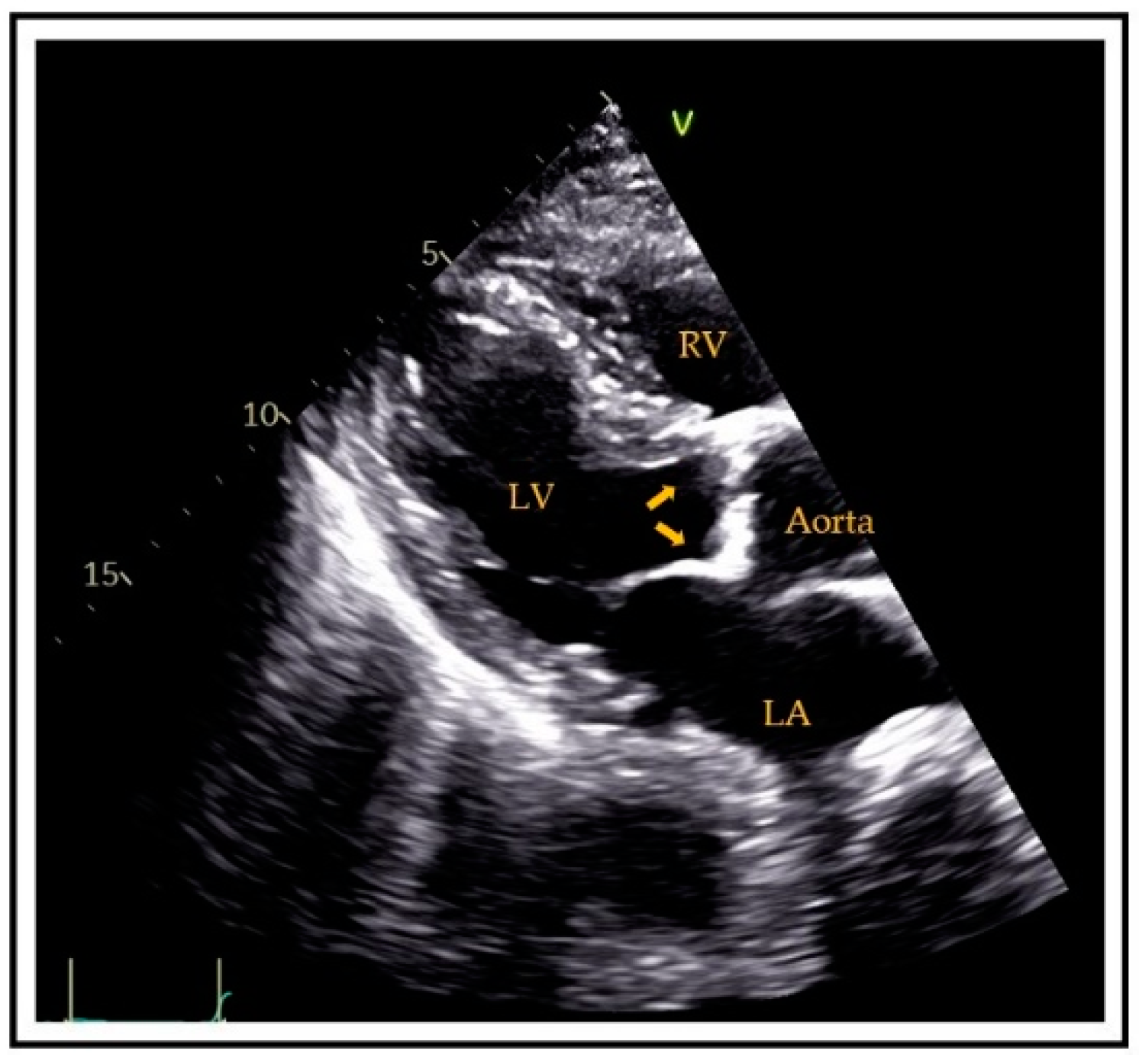
JCM | Free Full-Text | Cardiovascular Calcification as a Marker of  Increased Cardiovascular Risk and a Surrogate for Subclinical  Atherosclerosis: Role of Echocardiography
JCM | Free Full-Text | Cardiovascular Calcification as a Marker of Increased Cardiovascular Risk and a Surrogate for Subclinical Atherosclerosis: Role of Echocardiography

UOTW #57 Answer – Core Ultrasound

Anatomical and physiological complications related to left ventricular  apical aneurysm - Stoodley - 2017 - Sonography - Wiley Online Library
Anatomical and physiological complications related to left ventricular apical aneurysm - Stoodley - 2017 - Sonography - Wiley Online Library

Congenital left ventricular aneurysm of interventricular septum: prenatal  diagnosis and long-term management | Cardiology in the Young | Cambridge  Core
Congenital left ventricular aneurysm of interventricular septum: prenatal diagnosis and long-term management | Cardiology in the Young | Cambridge Core

Unruptured giant left ventricular pseudoaneurysm after silent myocardial  infarction | BMJ Case Reports
Unruptured giant left ventricular pseudoaneurysm after silent myocardial infarction | BMJ Case Reports


Echocardiographic assessment of left ventricular function recovery post  percutaneous coronary intervention of chronic total occlusions in patients  with low and mid‐range left ventricular ejection fractions - Samy - 2020 -  Echocardiography - Wiley ...
Echocardiographic assessment of left ventricular function recovery post percutaneous coronary intervention of chronic total occlusions in patients with low and mid‐range left ventricular ejection fractions - Samy - 2020 - Echocardiography - Wiley ...